First thing first, we went through previous results. We have a lot to go through and explain. First, there are the ones that are testing for extracellular enzymes which are starch, lipid, casein, and gelatin.
Starch Hydrolysis Test
First thing first, we analyzed our starch hydrolysis test. After gathering the plate, we poured grams iodine over it. Iodine only reacts with the starch and will form a glowing yellow shade around the bacteria, if the bacteria digested the carboyhydrates in the medium. Ours was negative, while our partner's group was slightly glowing, showing that theirs used starch a little bit.
First thing first, we analyzed our starch hydrolysis test. After gathering the plate, we poured grams iodine over it. Iodine only reacts with the starch and will form a glowing yellow shade around the bacteria, if the bacteria digested the carboyhydrates in the medium. Ours was negative, while our partner's group was slightly glowing, showing that theirs used starch a little bit.
The enzyme amalyse breaks starch into maltose which can be further
broken down into glucose. If the iodine reacts with starch, it produces a blue/black
color. If the bacteria has used amylase to break down the starch, clear zones appear around the bacteria and indicates a positive test.
 |
| Starch Hydrolysis: On left. |
Casein Hydrolysis
Next we looked at the casein hydrolysis. IF there is a clear zone around our enzyme, then it means that our bacteria can hydrolyze the protein, meaning that is secretes caseinase. Again, ours came back as a negative result while our partners came back as positive.
If the milk plate has a zone devoid of casein, there is a clear zone denoting the use of casein.
Gelatin Hydrolysis Test
Next we looked at the casein hydrolysis. IF there is a clear zone around our enzyme, then it means that our bacteria can hydrolyze the protein, meaning that is secretes caseinase. Again, ours came back as a negative result while our partners came back as positive.
If the milk plate has a zone devoid of casein, there is a clear zone denoting the use of casein.
 |
| Casein Hydrolysis: On left. |
Gelatin Hydrolysis Test
The gelatin hydrolysis test was used to determine if our bacteria could
digest gelatin. If there is liquid in the gelatin test tube, then we should keep
it in the refrigerator for 10-15 minutes to see if it is still liquid, which would indicate a positive test. Undigested gelatin, is a negative result.
Our bacteria broke down gelatin into short peptides and amino acids, and liquid was exhibited floating on top of the undigested gelatin in room temperature and after refrigeration (positive for the extracellular enzyme gelatinase).
 |
| Gelatin Test |
The spirit blue for ours did nothing. Our partner's turned slight
color but ours had no color change zone.
It was positive if it had the clear zone and ours did not. This means, that our bacteria did not have the extracellular enzymes called lipases; we had a negative result in this test.
 |
| Fat hydrolysis test: on left. |
Carbohydrate Fermentation
Intracellular enzymes: to determine their presence or absense we looked for any bubbles and any color changes-
no gas and no color change = negative,
color change = positive (whether gas or no gas)
1. Our sucrose was acidic, yellow, and contained no carbon dioxide – no
bubbles.
 |
| Sucrose |
2. Our glucose was acidic, yellow, and had a single bubble on the top of the tube; this means that carbon dioxide formation took place
 |
| Glucose |
3. The maltose was acidic, yellow, and also had one bubble, meaning that
fermentation again took place
 |
| Maltose |
4. Lastly, the lactose was a slight pink color with some bubbles at
the top, however we interpreted this as no fermentation and therefore a negative test result
MR Test
 |
| Lactose |
MR Test
Next we looked at the methyl red (MR) experiment. We used the test
tube that we began yesterday and split it into two test tubes (one for the MR test, and the other for the VP test). To the
first test tube, we added 5 drops of methyl red; if this kept the
red color, then it was a positive result for mixed fermentation. Ours was
negative.
The red color means that glucose was used and acetic, succinic, and formic acid was produced. If bacteria grows in glucose, it is likely to be positive here as was true with our bacteria.
VP TestThe red color means that glucose was used and acetic, succinic, and formic acid was produced. If bacteria grows in glucose, it is likely to be positive here as was true with our bacteria.
 |
| Methyl Red |
Next, we looked at the Voges-Proskauer (VP) test. To the second
test tube, we added 15 drops Barritt's reagent A and 5 drops of Barritt's
reagent B. We had to shake the bottom of the tube vigorously so that oxygen
permeated throughout the whole broth culture. We let this sit for 15 minutes, shaking it every so often. The end result was a pinkish color that was sustained after 10 minutes
of standing still.This pink color indicates that the bacteria produced butanediol
as one of the end products, by digesting the glucose present in the test tube.
 |
| Voges Proskeur Test |
TSIA Test
Next we did the TSIA test. We were looking for acidity of the slant and the butt, and then for gas. Red meant alkaline, and yellow meant acidic. If it had a dark black then hydrogen sulfide gas was produced. If the bacteria used glucose and produces acid, then you should see yellow color. It might use glucose is both colors, on the surfact the acid is able to be broken down while the one below can’t be.Ours was gaseous as well as yellow in the slant and yellow in the butt; the yellow-yellow means that it did use glucose. Glucose is only 0.1% of the media. Present in high concentration, acid is maintained both on top and bottom. Even on the top, we had enough sugar (sucrose and lactose) to produce acid.
Citrate Utilization Test
Next we did the TSIA test. We were looking for acidity of the slant and the butt, and then for gas. Red meant alkaline, and yellow meant acidic. If it had a dark black then hydrogen sulfide gas was produced. If the bacteria used glucose and produces acid, then you should see yellow color. It might use glucose is both colors, on the surfact the acid is able to be broken down while the one below can’t be.Ours was gaseous as well as yellow in the slant and yellow in the butt; the yellow-yellow means that it did use glucose. Glucose is only 0.1% of the media. Present in high concentration, acid is maintained both on top and bottom. Even on the top, we had enough sugar (sucrose and lactose) to produce acid.
 |
| TSIA Test |
Citrate Utilization Test
The Citrate Utilization Test was a test for bacterial growth where
the bacterium utilizes citrate as its sole source of carbon and energy. If dark
blue, as opposed to dark green appears on the slant, then the test is positive. Our agar slant turned blue
on top. Bacteria produces blue because it produces pyruvate and carbon dioxide.
Litmuse Milk
 |
| Citrate Test |
Our final test was the Litmus Milk. It was a test to see a plethora of different things aboutour bacteria.
a) If lactose is used and broken down to lactic acid, the litmus milk will turn pink. Ours stayed purple.
b) If gas formed, you would see the separation of the curd, which we did not.
c) If the litmus acts as an electron receptor for an anaerobic bacteria during anaerobic respiration, the litmus milk would turn into a white color. Ours did not, mean that it was aerobic.
d) If there was curd formation, then casein precipitated because of lactic acid being formed. A clot will be hard and will not contract. Ours was a hard clot.
e) If casein performed protein lysation, there would be ammonia as an end product which is alkaline and will turn the litmus a deep purple at hte top and a slight brown at the bottom. Our bacteria did not do this.
Our bacteria in the end was found to be nonlactose forming, non curdforming, clot forming aerobic bacteria with no protein lysation.
 |
| Litmus Milk Test |
Indole
If you see a red reagent color after the Kovac’s reagent was added to the cultured broth, it means that indole positive. The tryptophan was used by bacteria, created indole, and with Kovac’s reagent (wearing gloves, becuase it is a carcinogen) produces color. Ours was brown-red on top, and was negative for the indole test (did not produce indole).
If you see a red reagent color after the Kovac’s reagent was added to the cultured broth, it means that indole positive. The tryptophan was used by bacteria, created indole, and with Kovac’s reagent (wearing gloves, becuase it is a carcinogen) produces color. Ours was brown-red on top, and was negative for the indole test (did not produce indole).
 |
| Indole test |
New Test Results
Selective and differential medias
MacConkey – lactose – allows us to grow gram negative – the crystal
violet inhibits gram positive organism. In our results, our gram negative did
grow, proving that it is gram negative. If it did release acid, then the medium
surrounding the growth would be red (becuase the acids produced would change the pH and therefore the color of the medium), but ours was a negative test for fermentation. If
this was positive, it would have been a lactose fermenter. All Coliforms will
produce lactic acid from fermenting the lactose. This is a test that will be
used to test for E. coli in the water; ours therefore is also not a coliform.
 |
| MacConkey Agar: On left. |
EMB – Our test was a negative, it did not produce a green
sheen (if it did, then it would have hydrolyzed lactose, and created a bunch of acids to change the pH). This goes along with the results that we had received that it did not
ferment lactose.
 |
| EMB test: On right. |
Mannitol salt – Since 7.5% sodium chloride is in this medium, this media is
selective, and also has a differential fucntion because of the mannitol – separate those
that use mannitol and those that don’t. We do not have a yellow zone, one surrounding the growth, would indicate the fermentation of mannitol. If it grows it means that it grows in the salt, however ours was negative for both tests.
 |
| Mannitol Salt: On bottom. |
PEA – this medium used only to isolate gram positive
organisms; it is inhibitory to gram negative organisms, and therefore ours was a negative test. Our bacteria did not even grow in this agar medium.
 |
| PEA test: On bottom. |
Blood agar – gamma – means no lysis, alpha = partial with greenish pigment, beta = complete - some lysis but not completely clear. Our result is
gamma; there was no lysis of red blood cell.
 |
| Blood agar test: on right. |
All the above new tests – selective and differential mediums.
Nitrate – This test tries to see if our bacteria use nitrate, NO3, and
converts it straight into nitrogen gas or ammonia. In this experiment, we have two reagents to add, Sulfanithic acid and napththylamine.If these two drugs create no red color – possibility nirtates not used at all, or the nitrites have
become ammonia – the first sulfanilic acid is just to test nitrites. You have
to go back and see if nitrates are still there by adding zinc powder to possibly reduce the nitrates left in solution; if they are, then the bacteria did not
use the nitrates at all and adding zinc will make the solution red, showing a false positive. If they are not there, then they went up into ammonia. Ours did not
have gas in the tube. The second test – the zinc converts the nitrates to
nitrites. If you see red after zinc, then it means the nitrates are there and
they were never used. Ours, after the add of the sulfinilic acid and alpha
naphtthylamin became a deep dark red, meaning that it created the nitrite in
it.
 |
| Nitrate Test |
Urea – This test looks to see if our bacteria can use urea; if it has urease
it can produce carbon dioxide, water, and ammonium. Ammonia is alkaline – so the
phenyl red will turn into pink. Ours was yellow in color, meaning that it did
not use urea.
After completing all of these test, we got to play detectve and find out what our bacteria was. By looking online and by first using the shape and the gram staining, we finally located it. It was Serratia Marcescens. This is the list of its properties, and how ours matched up with it.
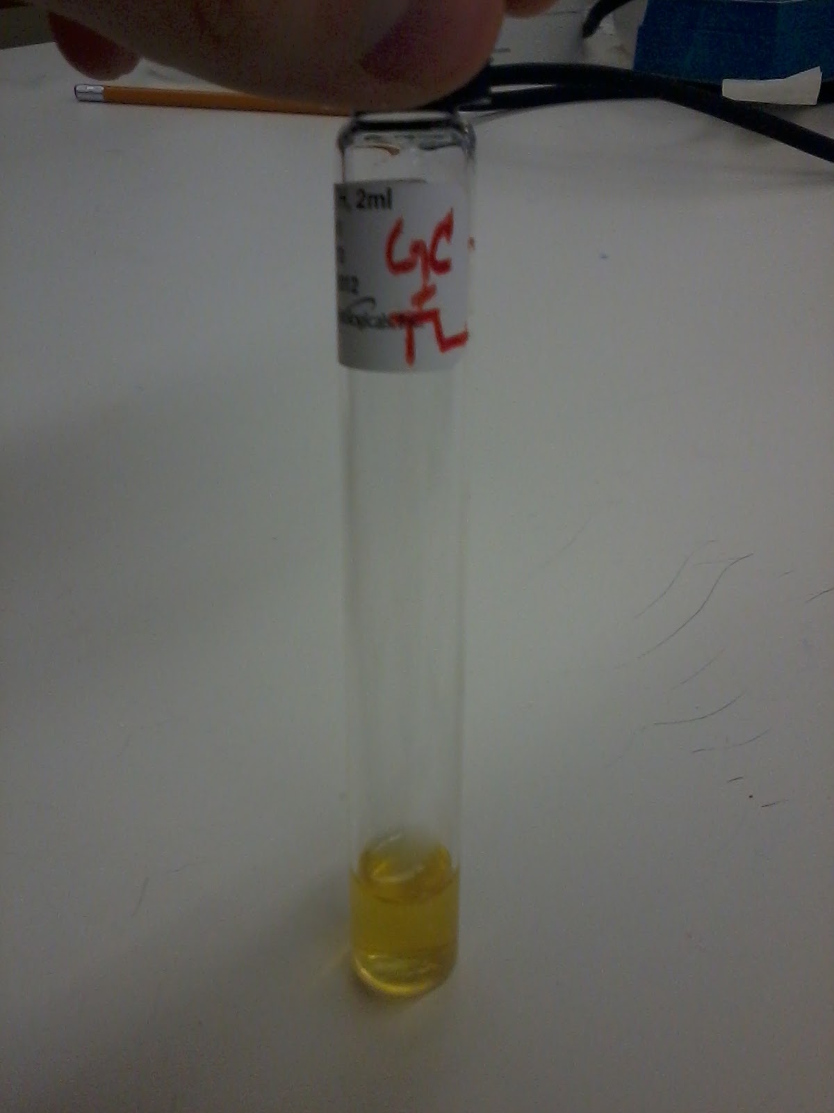 |
| Urea Test |
| Test | Result |
|---|---|
| Gram stain | - |
| Oxidase | - |
| Indole production | - |
| Methyl Red | >70% - |
| Voges-Proskaeur | + |
| Citrate (Simmons) | + |
| Hydrogen sulfide production | - |
| Urea hydrolysis | >70% - |
| Phenylalanine deaminase | - |
| Lysine decarboxylase | + |
| Motility | + |
| Gelatin hydrolysis, 22 C | + |
| Acid from lactose | - |
| Acid from glucose | + |
| Acid from maltose | + |
| Acid from mannitol | + |
| Acid from sucrose | + |
| Nitrate reduction | + (to nitrite) |
| Deoxyribonuclease, 25 C | + |
| Lipase | + |
| Pigment | some biovars produce red |
| Catalase production (24h) | + |
* App of the Day - Serratia Marcescens is commonly found in restrooms on the tiles, in the corners of shows, at the base of faucets. It can be difficult to get rid of, but use a bleach-based disinfectant, and it should come up. Serratia Marcescens can cause bacterial infection in the urinary tract, respitory tract, wounds, and the eye.
No comments:
Post a Comment