Saturday, June 2, 2012

Day 13 - Record Antibiotics and oils, Hamburger extract results, andHIV Test

First thing first, we checked out our results from yesterday. We gathered our two separate plates of oils and antibiotics and looked to see which inhibited growth. Taking a small ruler, we measure in millimeters how far the circle was clear of bacteria around the disk of the given solution. The larger the disk means the larger the antibacterial age power of the solution.

Top: Antibiotics, Bottom: Oil
Results:


Antibiotics

Oils

1. Penicillin
No inhibition
1. Clove
10 mm
2. Erythromycin
18 mm
2. Cinnamon
20 mm
3. Chloramphenicol
20 mm
3. Thyme
22 mm
4. Novobiocin
No inhibition
4. Eukalyptus
No inhibition
5. Augmentin
15mm
5. Ginger
No inhibition


The best that worked on our bacteria out of the tests was thyme, with Cinnamon and Chloramphenicol coming in second. Over all, the oils had a better inhibition of the bacteria than the antibiotics. Our gram negative bacteria showed similar results to others in the class who also had gram negative bacteria. Out of the oils, in general, the thyme, cinnamon, and gloves tended to work best on gram negative. For the antibiotics, the chloramphenicol worked the best. Again, this worked in general for gram negative bacteria because it bypassed other systems and attacked the protein synthesis.

We also checked our results from the Hamburger extract experiment. What was an indication of the antibody reaction was a small crescent shaped cloud in between where the hamburger extract was and the anti solutions surround it. The only antibody that was formed in our result was the anti-bovine. This means that cow meat was really the only meat used in this hamburger.

Crescent seen between top right hole and middle hole. 

Next, we split the class into two groups and did the Elisa test.

Elisa test setup.

This is a test to see if there is HIV the system of a patient. This is done by detecting the antibodies for HIV in a blood sample. The basic concept of this test is that if there is HIV in the system, the body will have formed the antibodies which are meant to attach to the antigens of the HIV. We separate out this antigen, attach it to the side wall of a tube, then we add the blood. If there is antibodies in it, it will bind to the antigen, if not, then it will be washed away. To determine if the blood same left antibodies we add an animal (goat) antibody. The goat antibody is an antibody to the HIV antibody in humans. If the HIV antibody is there, the goat antibody will bind. If not, it will be washed away. This second antibody can be detected because it has an enzyme connected to it that will change color in the presence of a substrate.

Therefore, if the patient's sample is HIV positive, the human antibody will bind to the antigen, the goat antibody with the enzyme will bind to the human antibody, and the substrate will cause the solution to turn blue. If the patien'ts sample is HIV negative, there will be no antibody to attach to the antigen, and therefore not human antibody for the goat antibody with the enzyme to attach to, and everything will be washed away, leaving the solution in the tube clear of color.

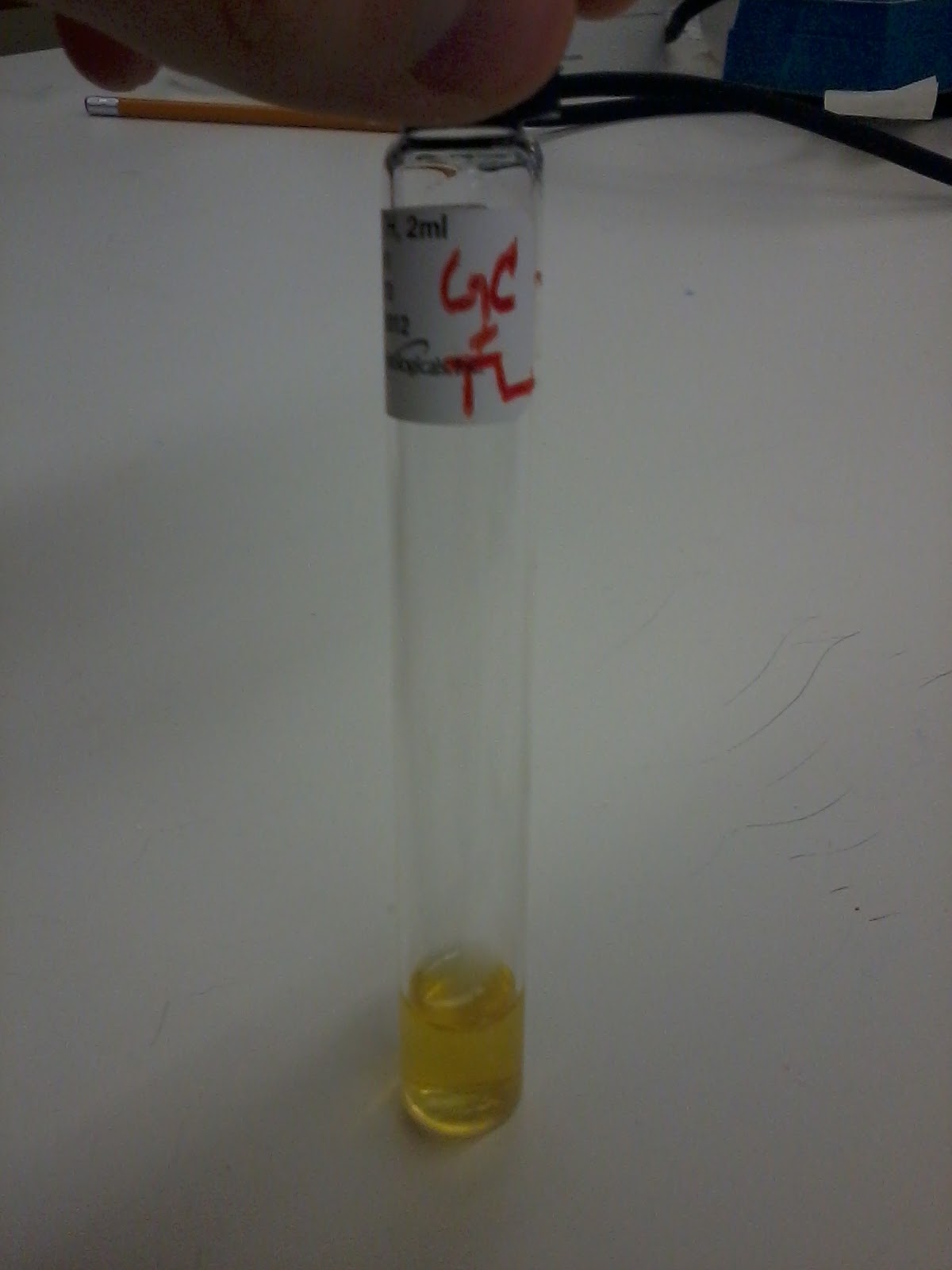
For our test in particular, we had a set of 12 small tubes in a well tray. In the first 3, we added a negative sample. In the second, a positive one, and in the last the variable sample that we are trying to figure out. The positive should turn blue, the negative stay clear, and whichever the variable will look like. Our test failed and all stayed clear. The other group, besides a problem in two of the tubes, proved that their sample was HIV positive.

Other teams positive HIV test. 


*App of the day - Dr. Joseph is doing an experiment, looking into the affects of cinnamon. The oils have great antibacterial power, but a lot have adverse side effects. Some will kill red blood cells, which is a major problem. Cinnamon was found to be a good middle ground, with antibacterial power as well as the least affect on red blood cells. To use on the skin, there is no problem will cinnamon oil. Ingesting straight cinnamon oil, might be dangerous. But if you slightly increase cinnamon in your diet, it might keep bacteria in line. 

Day 12 - Antibiotic Test and Hamburger Extract

Antibiotic Test

First step first, we took a plate and streaked it all over with our bacteria. We then added the antibiotic disks over the streak plate, over the numbers we had written on the underside of the plate. If the discs secret antibiotics, then bacteria around it will not grow and make a clear zone where bacterial colonies are killed; you won't see bacteria growing around disk. The larger clear zone, the better the antibiotic.
To put the disks on, you take the 70% ethanol, and dip the forceps in it. You then burn the alcohol off the forceps, take the sample antibiotic out of the package, and lay it on the dish over its corresponding number from below:
Antibiotic set up


Antibiotics:
1. Penicillin
2. Erythromycin
3. Chloramphenicol
4. Novobiocin
5. Augmentin - very old

Oils Test

To see if they have any antibiotic affect, we used the same technique as above. 5 microliters of oil were deposited onto a sterile piece of paper, after the forceps had been sterilized. Each sterile piece of paper was put over the corresponding number below:

All of the oils.


Oils:
1.Clove
2. Cinnamon
3.Thyme
4. Eucalyptis
5. Ginger

Oil and Antibiotics, ready to go into incubator.
Hamburger Extract Test

The way antibodies work is that each antibody can bind with two antigens. Antibodies bind only with specific antigens. This test determines whether the protein in the middle well is bovine (cows) albumin or if it has been contaminated with other meats. Albumin is in all blood and maintains osmotic potential. The way to make an anti-horse bovine is to add a horse albumin to a goat. The goat's system will see this as foreign and will form antibodies against this. We use this to detect the horse albumin, and therefore horse meat.

To determine if the hamburger extract has contamination with other meats, one group does template II and the other template III. We were told to do the hamburger extract test (template I), to see if the hamburger was pure. First, we had to make four holes in the agar using a dropper to suck up the agar, and pulling it out to make the wells. After the 4 holes are made in the pattern assigned, we put hamburger extract, goat anti-horse albumin, goat anti-bovine albumin, and goat anti-swine albumin, each in one of the four wells, adding one drop of each to its corresponding hole, and wait until tomorrow to see the results (this test did not have to be incubated).

Plate used for Hamburger Extract


*App of the Day - Our test was found to be pure hamburger meat. It was free from other contamination. But, that hamburger meat may be full of other things that we don't even realize. Nitrate is one of the preservatives often used in meat. It also might be interesting to test a hot dog with this method. Hmm.. maybe some things are better off not known.

Day 11 - Explanations of Old and Viewing of New

Previous Results


First thing first, we went through previous results. We have a lot to go through and explain. First, there are the ones that are testing for extracellular enzymes which are starch, lipid, casein, and gelatin.

Starch Hydrolysis Test


     First thing first, we analyzed our starch hydrolysis test. After gathering the plate, we poured grams iodine over it. Iodine only reacts with the starch and will form a glowing yellow shade around the bacteria, if the bacteria digested the carboyhydrates in the medium. Ours was negative, while our partner's group was slightly glowing, showing that theirs used starch a little bit.
The enzyme amalyse breaks starch into maltose which can be further broken down into glucose. If the iodine reacts with starch, it produces a blue/black color. If the bacteria has used amylase to break down the starch, clear zones appear around the bacteria and indicates a positive test.
Starch Hydrolysis: On left.


Casein Hydrolysis


     Next we looked at the casein hydrolysis. IF there is a clear zone around our enzyme, then it means that our bacteria can hydrolyze the protein, meaning that is secretes caseinase. Again, ours came back as a negative result while our partners came back as positive.
If the milk plate has a zone devoid of casein, there is a clear zone denoting the use of casein.
Casein Hydrolysis: On left.


Gelatin Hydrolysis Test

     The gelatin hydrolysis test was used to determine if our bacteria could digest gelatin. If there is liquid in the gelatin test tube, then we should keep it in the refrigerator for 10-15 minutes to see if it is still liquid, which would indicate a positive test. Undigested gelatin, is a negative result.
Our bacteria broke down gelatin into short peptides and amino acids, and liquid was exhibited floating on top of the undigested gelatin in room temperature and after refrigeration (positive for the extracellular enzyme gelatinase).

Gelatin Test
Fat Hydrolysis Test

     The spirit blue for ours did nothing. Our partner's turned slight color but ours had  no color change zone. It was positive if it had the clear zone and ours did not. This means, that our bacteria did not have the extracellular enzymes called lipases; we had a negative result in this test.
Fat hydrolysis test: on left.


Carbohydrate Fermentation


     Intracellular enzymes: to determine their presence or absense we looked for any bubbles and any color changes-
    no gas and no color change = negative,
    color change = positive (whether gas or no gas)
1. Our sucrose was acidic, yellow, and contained no carbon dioxide – no bubbles.


Sucrose

2. Our glucose was acidic, yellow, and had a single bubble on the top of the tube; this means that carbon dioxide formation took place


Glucose

3. The maltose was acidic, yellow, and also had one bubble, meaning that fermentation again took place


Maltose

4. Lastly, the lactose was a slight pink color with some bubbles at the top, however we interpreted this as no fermentation and therefore a negative test result
Lactose


MR Test

     Next we looked at the methyl red (MR) experiment. We used the test tube that we began yesterday and split it into two test tubes (one for the MR test, and the other for the VP test).  To the first test tube, we added 5 drops of methyl red; if this kept the red color, then it was a positive result for mixed fermentation. Ours was negative.
The red color means that glucose was used and acetic, succinic, and formic acid was produced. If bacteria grows in glucose, it is likely to be positive here as was true with our bacteria.
Methyl Red


VP Test
     Next, we looked at the Voges-Proskauer (VP) test. To the second test tube, we added 15 drops Barritt's reagent A and 5 drops of Barritt's reagent B. We had to shake the bottom of the tube vigorously so that oxygen permeated throughout the whole broth culture. We let this sit for 15 minutes, shaking it every so often. The end result was a pinkish color that was sustained after 10 minutes of standing still.This pink color indicates that the bacteria produced butanediol as one of the end products, by digesting the glucose present in the test tube.


Voges Proskeur Test


TSIA Test


Next we did the TSIA test. We were looking for acidity of the slant and the butt, and then for gas. Red meant alkaline, and yellow meant acidic. If it had a dark black then hydrogen sulfide gas was produced.   If the bacteria used glucose and produces acid, then you should see yellow color. It might use glucose is both colors, on the surfact the acid is  able to be broken down while the one below can’t be.Ours was gaseous as well as yellow in the slant and yellow in the butt; the yellow-yellow means that it did use glucose. Glucose is only 0.1% of the media. Present in high concentration, acid is maintained both on top and bottom. Even on the top, we had enough sugar (sucrose and lactose) to produce acid.


TSIA Test


Citrate Utilization Test
The Citrate Utilization Test was a test for bacterial growth where the bacterium utilizes citrate as its sole source of carbon and energy. If dark blue, as opposed to dark green appears on the slant, then the test is positive. Our agar slant turned blue on top. Bacteria produces blue because it produces pyruvate and carbon dioxide.


Citrate Test
Litmuse Milk

Our final test was the Litmus Milk. It was a test to see a plethora of different things aboutour bacteria.
   a) If lactose is used and broken down to lactic acid, the litmus milk will turn pink. Ours stayed purple.
   b) If gas formed, you would see the separation of the curd, which we did not.
   c) If the litmus acts as an electron receptor for an anaerobic bacteria during anaerobic respiration, the litmus milk would turn into a white color. Ours did not, mean that it was aerobic.
   d) If there was curd formation, then casein precipitated because of lactic acid being formed. A clot will be hard and will not contract. Ours was a hard clot.
   e) If casein performed protein lysation, there would be ammonia as an end product which is alkaline and will turn the litmus a deep purple at hte top and a slight brown at the bottom. Our bacteria did not do this.

Our bacteria in the end was found to be nonlactose forming, non curdforming, clot forming  aerobic bacteria with no protein lysation. 


Litmus Milk Test


Indole
     If you see a red reagent color after the Kovac’s reagent was added to the cultured broth, it means that indole positive. The tryptophan was used by bacteria, created indole, and with Kovac’s reagent (wearing gloves, becuase it is a carcinogen) produces color. Ours was brown-red on top, and was negative for the indole test (did not produce indole).


Indole test


New Test Results


Selective and differential medias


MacConkey – lactose – allows us to grow gram negative – the crystal violet inhibits gram positive organism. In our results, our gram negative did grow, proving that it is gram negative. If it did release acid, then the medium surrounding the growth would be red (becuase the acids produced would change the pH and therefore the color of the medium), but ours was a negative test for fermentation. If this was positive, it would have been a lactose fermenter. All Coliforms will produce lactic acid from fermenting the lactose. This is a test that will be used to test for E. coli in the water; ours therefore is also not a coliform.
MacConkey Agar: On left.


EMB – Our test was a negative, it did not produce a green sheen (if it did, then it would have hydrolyzed lactose, and created a bunch of acids to change the pH). This goes along with the results that we had received that it did not ferment lactose.

EMB test: On right.


Mannitol salt –  Since 7.5% sodium chloride is in this medium, this media is selective, and also has a differential fucntion because of the mannitol – separate those that use mannitol and those that don’t. We do not have a yellow zone, one surrounding the growth, would indicate the fermentation of mannitol. If it grows it means that it grows in the salt, however ours was negative for both tests.

Mannitol Salt: On bottom.

PEA – this medium used only to isolate gram positive organisms; it is inhibitory to gram negative organisms, and therefore ours was a negative test. Our bacteria did not even grow in this agar medium.

PEA test: On bottom.

Blood agar – gamma – means no lysis, alpha = partial with greenish pigment, beta = complete  - some lysis but not completely clear. Our result is gamma; there was no lysis of red blood cell.
Blood agar test: on right.


All the above new tests – selective and differential mediums.

Nitrate – This test tries to see if our bacteria use nitrate, NO3, and converts it straight into nitrogen gas  or ammonia. In this experiment, we have two reagents to add, Sulfanithic acid and napththylamine.If these two drugs create no red color – possibility nirtates not used at all, or the nitrites have become ammonia – the first sulfanilic acid is just to test nitrites. You have to go back and see if nitrates are still there by adding zinc powder to possibly reduce the nitrates left in solution; if they are, then the bacteria did not use the nitrates at all and adding zinc will make the solution red, showing a false positive. If they are not there, then they went up into ammonia. Ours did not have gas in the tube. The second test – the zinc converts the nitrates to nitrites. If you see red after zinc, then it means the nitrates are there and they were never used. Ours, after the add of the sulfinilic acid and alpha naphtthylamin became a deep dark red, meaning that it created the nitrite in it.
Nitrate Test

Urea – This test looks to see if our bacteria can use urea; if it has urease it can produce carbon dioxide, water, and ammonium. Ammonia is alkaline – so the phenyl red will turn into pink. Ours was yellow in color, meaning that it did not use urea.

Urea Test
After completing all of these test, we got to play detectve and find out what our bacteria was. By looking online and by first using the shape and the gram staining, we finally located it. It was Serratia Marcescens. This is the list of its properties, and how ours matched up with it. 

TestResult
Gram stain-
Oxidase-
Indole production-
Methyl Red>70% -
Voges-Proskaeur+
Citrate (Simmons)+
Hydrogen sulfide production-
Urea hydrolysis>70% -
Phenylalanine deaminase-
Lysine decarboxylase+
Motility+
Gelatin hydrolysis, 22 C+
Acid from lactose-
Acid from glucose+
Acid from maltose+
Acid from mannitol+
Acid from sucrose+
Nitrate reduction+ (to nitrite)
Deoxyribonuclease, 25 C+
Lipase+
Pigmentsome biovars produce red
Catalase production (24h)+

* App of the Day - Serratia Marcescens is commonly found in restrooms on the tiles, in the corners of shows, at the base of faucets. It can be difficult to get rid of, but use a bleach-based disinfectant, and it should come up. Serratia Marcescens can cause bacterial infection in the urinary tract, respitory tract, wounds, and the eye. 

Friday, June 1, 2012

Day 10 - Urea, Differential Plates, PEA, NItrate, Oxidase, and Yogurt

Urea Hydrolysis Test

     This test determines the ability of a bacterium to hydrolyze urea with an enzyme called urease; urea is a small molecule excreted in urine to elimniate nitrogenous waste from the body. This plate contains a pH indicator also called phenol red, because urease breaks urea down into carbon dioxide and ammonia which makes the pH more alkaline. So, a positive test makes the test tube a pink color, while a yellow color indicates no reaction (a negative result).

Urea test: before incubation.


Differential Plates
    a) MacConkey
          This selective and differential media, differentiates among gram-negative enteric bacilli, based on their ability to grow on the medium and ferment lactose; these agar plates are often used in to process fecal matter specimens. Gram positive bacteria are inhibited on this growth plate, because crystal violet and bile salts are included in the medium. Lactose-fermenting colonies produce acids and have a pink to red color becuase of the pH indicator; non-lactose-fermenting colonies are colorless.
McConkey agar before incubation: Our test on left side. 


    b) EMB - Eosin Methylene Blue Plate
          This test isolates and differentiates gram-negative enteric bacilli. It contains eosin dye, methylene dye, lactose, and sucrose.  It is a differential media that shows what bacteria ferment lactose and/or sucrose, and those that do not. If a bacteria does ferment these sugars, the dyes will precipitate on the colonies becuase of lower pH; slight carbohydrate fermentation causes a purple/pinkish sheen to the bacterial colonies and no fermentation causes no change in colony formation or color of the agar.
EMB before incubation: Our test on left side.


    c) Mannitol Salt
          Manitol salt agar is meant to be a selective media to islate bacteria based ont heir salt tolerance and differentaite among these isolates for mannitol fermentation. Acid prodcued from the fermentation will cause the pH indicator, phenol red, to change to a yellow colored zone around the bacterial colonies (positive test); a negative test results in no change of color around bacterial colonies.


    d) Blood Agar
          This plate is an enriched medium with 5% red blood cells, to isolate and grow fastidious bacteria in clinical specimens. This differential media separates bacteria according to their ability to lyse blood cells- alpha, beta, or gama hemolysis. Alpha hemolysis gives off a green sheen to the agar plate, while in beta hemolysis, clear areas can be seen around bacterial colonies. Gama hemolysis shows no change in the blood agar plate.


Phenylethyl Alcohol Agar
     This growth plate isolates gram-positive bacteria from a specimen containg a mixture of gram-positive and gram-negative bacteria. It is a selective media, becuase PEA inhibits or slows the growth of gram-negative gacteria by interfering with DNA synthesis.

PEA agar before incubation: Our test on left.


Nitrate Reduction Test
     The nitrate test determines if a bacterium is able to reduce nitrate ions ot either nigrate ions or nitrogen gas. When the enzyme nitrate reductase is present, it facilitates the recution of the nitrate ion to nitrite ions and water. Anaerobic bacteria are able to reduce nitrate completely to molecular nitrogen in a process called denitrification. After inoculation and incubation, reagents sulfanilic acid and dimethyl-a-naphthylamine are added and if nitrite is present (a positive test) the solution will turn pink; if not, the solution will stay a yellowish color. The addition of zinc powder may be added becuase it will reduce nitrate ions to nitrite ions- this would be a false positive test.

Nitrate test before incubation.


Oxidase Test
     This test determines if bacteria have cytochrome oxidase, a necessary enzyme in the electron transport chain during respiration. We did not follow this procedure completely in that we did not culture a plate, but we used an already cultured plate to test. In this test a reagent N,N,N,N, tetramethy-p-phenylenediamine is added. If the color of the bacteria immediately turned purple, the test was positve for cytorchrome oxidase, if not the color stayed the same (negative test).

Since we already has a cultured plate, we added the reagent and at first it was difficult to tell if our bacteria would change color (especially since it was already a deep red color). After comparing it to others' tests, our oxidase test came out negative and did not turn purple.

Oxidase test: Top.

Making Yogurt
To make yogurt you heat up milk (preferably whole milk) by heating it until it boils and puffs up with bubbles; you do this to sterilize the milk, and give the bacteria you add to it, a broth to grow in. You then and cool it down to 37 degree centigrad (which makes the best yogurt) and add the active yogurt cultures and put it into 37 incubator (this can also be done in an oven overnight, with just the oven light on). To cool faster, transfer the milk from one cup to another cups, allowing the milk to airate. At home, if you touch the mug, it should be almost your temperature. We sprayed all utensiles with alcohol to keep them sterile, and from contaminating the milk with other unwanted bacteria. We made two different kinds of yogurt; one with the kroger active cultures and one with Dr. Joseph's cultures, originally from India. For them to be good active cultures, they should form  yogurt within 6-24 hours.


We then finished our last movie, Outbreak. :)

Day 9 - Outbreak

On this day in lab, we began to watch Outbreak. This is a movie made in 1995 that shows how  a virus spreads. We got about half-way through the movie and will finish it at another time.


*App of the Day - One must always be careful not to spread any bacteria and must especially be conscious of contact with animals. In the movie, the virus is spread through monkey sputum, sharing food, and kissing someone who is sick; disease can be spread through contact, droplet, or airborne means. You have to be aware of the possibility of sickness becuase otherwise, especially working as a nurse, one could endanger the lives of patients by careless behavior.

Day 8 - Recording Results


Today, we recorded the results for all of the tubes and plates that we began on the previous day. Dr. Joseph did not explain the results fully yet, and we will put up all of the results, along with the descriptions, at a later date.

Our full collection of tubes and plates. 
* App of the Day - Lactic acid fermentation is one way milk ferments into a 'sour' curd. This is a sign that milk is no longer good to drink


Day 7 - Starch, Casein, Gelatin, Carbohydrates, MRVP, Citrate, Indole, Litmus Milk, and TSI Tests


Before starting this, we checked out our anaerobic tests. These tests for some reason did not work out. The methylene blue strip was still blue, indicating the presence of oxygen. In order to still use these plates we prepared (that in the end had grown in the aerobic conditions), we tested this with hydrogen peroxide. If it formed bubbles, then it produced carbon dioxide as a result of the catalase enzyme breaking down the hydrogen peroxide. If there was no bubbling, then it produced no carbon dioxide. Ours produced a large amount of bubbles, while our partner's group did not produce any.

Our sample, upper right, large amounts of bubbles.

Today began a very fast-paced day of inoculating tubes and spreading bacteria on plates. We prepared the tubes and plates for 14 different tests.


1.) Starch Hydrolysis Test

     In this test, we are looking to see if our bacteria hydrolyzes/digests starch by the extracellular enzyme called amylase. This enxyme breaks the bonds that link the glucose subunits. If a bacteria has this enzyme, it will have glucose and maltose as biproducts, creating a large growth of colonies on this plate. Using aseptic technique, our pink bacteria was inoculated onto the slide and will incubate until tomorrow.


2.) Casein Hydrolysis Test

     Casein is the predominate protein in milk, and can be used as a substrate to assess the production for proteinases. In this experiment, we will determine wheter our bacteria has the ability to digest casein, by inoculating some of our bacteria onto a skim milk agar plate, streaking a short line on the agar surface, and will incubate until tomorrow. The products for a postive test are amino acids and peptides.

3.) Gelatin Hydrolysis Test

     Gelatin is a processed form of collagen, an important protein in connective tissue. When boiling the bones of cows and pigs, the product is gelatin, a semi-solid gel. This test distinguishes bacteria that have the enzyme gelatinase, which can break the peptide bonds in gelatin, causing the gel to liquify it into amino acides and short peptides. This procedure is slightly different than the other two above, in that this one calls for an inoculated needle to be stabbed into a gelatin test tube, to once again be incubated until later.

4.) Carbohydrates Fermentation


     The four carbohydrate tests below will determine whether our bacteria will ferment a particular carbohydrate. This test looks for two results: a possible color change of the phenol red added to the test tube (yellow means acidic, red means basic) and possible gas formation from the fermentation of the following carbohydrates. We inoculated the test tubes, and incubated them.

      a.) Glucose
      b.) Sucrose
      c.) Maltose
      d.) Lactose

5.) Methyl Red Test  

     This test looks for products of mixed-acid fermentation of glucose into significant amounts of organic acids (acetic, succinic, and formic acid as well as gas). The test tubes have a pH indicator in them called methyl red (hence the name of the test) that remains red in a positive test (acidic) and turns orange/yellow in a negative test (alkaline). We simply used aseptic technique again to inoculate the broth inside the test tube, and wait until tomorrow for results in the incubator.

6.) Voges-Proskauer Test

     Some bacteria have a 'butanediol fermentation pathway' utilized to ferment glucose. This test looks for the protein acetoin, a vital intermediate in this pathway. Organic acids are products of this pathway, and a red color denotes a positive test becuase the presence of oxygen gas and acetoin reacts with the VP reagent. No color change (yellow) is a negative test. We carefully inoculated the bacteria into the corresponding test tube, and put it into the incubator.

7.) Citrate Utilization Test

     This test looks for the membrane protein citrate permease; an enzyme that breaks citrate into pyruvate and carbon dioxide. This test determines if our bacteria can utilize citrate as the sole source of carbon. If the bacteria produces carbon dioxide, it will bind with the sodium and form sodium carbonate, an alkaline compound that changes the color of the pH indicator (bromothymol blue) blue from the initial green color. After inoculating the agar slant with our bacteria, we wait until tomorrow.

8.) Indole - Tryptophan Degradation - Test

     This test determines if a bacteria splits the amino acid tryptophan into indole and and pyretic acid. Though indole is a byproduct not used by the bacteria, it is the marker this test uses. If positive for indole, the bacteria will have broken tryptophan and oxygen into pyruvate, ammonia, and indole by the protein tryptophanase. We will know tomorrow if by inoculating our test tube, our bacteria uses trptophan as a source of endergy by degrading this amino acid. A red strip on the top of the broth tomorrow after added procedures will indicate a positive test for tryptophan use, and a negative test will show a yellow strip.

9.) Litmus Milk Test

     This test, differentiates between bacteria as to their ability to utilize lactose, protein, and litmus in litmus milk. Litmus here serves as a pH indicator, and turns pink in an acidic environment (lactose fermenting into acids), and blue in an alkaline environment. If the solution becomes too acidic, the casein in the milk may become denatured and from a firm curd; some bacteria may even turn the litmus clear. If casein is altered by another protein called rennin, a soft curd is formed. Proteolytic bacteria digest milk proteins into peptides and amino acids; all the above observations are important in this test and if casein is converted to amino acids by proteases, and to ammonia by deaminases, the whole solution will turn blue.  A lot will go on in the test tube overnight, but this is again just a simple inoculation- using aseptic technique of course... and we wait for tomorrow.

10.) TSIA Test

     This test differentiates among gram-negative enteric bacilli, Enterobacteriaceae, (which means that gram positive bacteria will not grow), as to their ability to ferment glucose, lactose, and sucrose, and to produce hydrogen disulfide. The triple sugar agar contains lactose, sucrose, and glucose and if our bacteria ferments any of them, the pH of the agar will drop considerably because of the organic acid byproducts. If the bacteria starts to ferment the protein in the medium instead of (or after) fermenting the sugars, the pH becomes alkaline and liberages ammonia from the amino acids. If acidic the medium will turn yellow; if alkaline, the medium will turn red. Black precipitates indicate hydrogen sulfide production, and gas indicates the production of ammonium gas. We inoculate the agar plate and place it in the incubator.


11.) Triglyceride (Fat) Hydrolysis Test

     This test differentiates bacteria that can break down lipids, which are primarily water-insoluble molecules. This test looks for bacteria that use the enzyme called lipase to break down the fat into fatty acids and glycerol resulting in a clear zone around the bacterial growth after incubation. Today we inoculated the triglyceride agar plate, and wait until tomorrow to determine the results.

*App of the Day - The reason why the dish bubbled when hydrogen peroxide was put on it was the same reason why wounds will bubble with the addition of Hydrogen peroxide. In either case, the H2O2 is being broken down by an enzyme (which in the body is catalase) to become H20 and O2. The bubble are actually bubbles of oxygen gas.